


Summary
- Microsoft Research introduced CARE-X, a chest X-ray reading system that combines flexible reasoning, calibrated predictions, and measurement-based tools
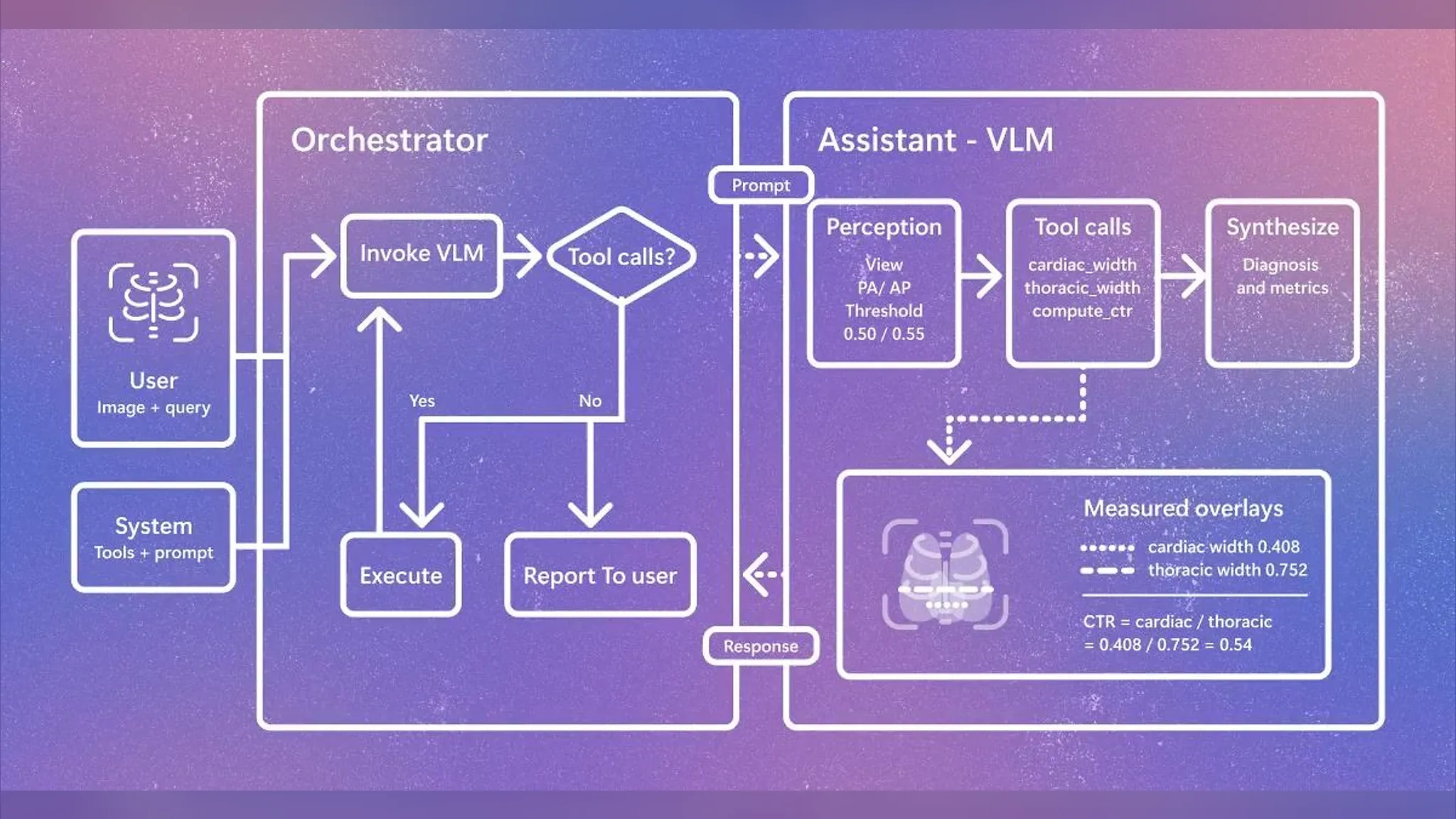
- The system works by having an orchestrator decide whether tool calls are needed, while a VLM (vision-language model) assistant measures cardiac width and thoracic width to calculate the cardiothoracic ratio (CTR)
- In the example image, a cardiac width of 0.408 and thoracic width of 0.752 were measured, yielding a CTR of 0.54 — a value that falls within the 0.50–0.55 clinical threshold range used to determine cardiomegaly
Two rulers drawn over an X-ray
A single diagram released by Microsoft Research shows two measurement lines overlaid on a chest X-ray image. One measures the widest point of the heart, the other the full width of the chest cavity. Dividing these two values yields the cardiothoracic ratio (CTR) — a long-standing clinical metric indicating the proportion of the chest cavity occupied by the heart, used to gauge whether the heart is abnormally enlarged. On August 11, Microsoft Research introduced a system applying this approach, called CARE-X, via its official X account.
In the example shared, a cardiac width of 0.408 and thoracic width of 0.752 were measured, yielding a CTR of 0.54. Clinically, a value above 0.5 is typically treated as suggestive of cardiomegaly, and this figure sits right at that boundary. Rather than delivering a diagnosis as a sentence alone, the system displays the underlying numbers on screen as supporting evidence.
Two layers of interpretation: orchestrator and VLM
CARE-X's structure is divided into two layers. When a user inputs an image and a question, an orchestrator receives it and passes a prompt to an assistant VLM (vision-language model — a model that processes images and text together). The assistant first perceives the imaging orientation (PA/AP, posteroanterior or anteroposterior), and if it determines that a tool call is necessary, it executes functions such as cardiac_width, thoracic_width, and compute_ctr. When these results return to the orchestrator, and no further tool calls are deemed necessary, the system compiles a final report and delivers it to the user.
| Stage | Handled by | Processing |
|---|---|---|
| Input | User | Image + query |
| Decision | Orchestrator | Calls VLM, branches on whether tools are needed |
| Perception | Assistant-VLM | Determines imaging view, applies 0.50/0.55 thresholds |
| Measurement | Tool calls | cardiac_width, thoracic_width, compute_ctr |
| Synthesis | Assistant-VLM | Presents diagnosis together with figures |
Organizing the measured values themselves into a table makes their distance from the threshold clear.
| Item | Value |
|---|---|
| Cardiac width | 0.408 |
| Thoracic width | 0.752 |
| Calculated CTR | 0.54 |
| Applied threshold | 0.50 / 0.55 |
From generated reports to verifiable evidence
Until now, most AI systems for chest X-rays worked by viewing an image and writing out a complete report in one pass. The problem is that it's difficult to trace back what evidence produced those sentences. What CARE-X emphasizes, as reflected in its description as a "measurement-based tool," is a structure that leaves behind calculable intermediate values before arriving at a diagnosis. This means a physician isn't limited to simply receiving a report and judging whether it's right or wrong — they can also check the concrete figures for cardiac and thoracic width along with the calculation process. Notably, the orchestrator itself decides whether a tool call is needed. Rather than forcing the same calculation on every image, this suggests the system embeds flexible reasoning that invokes measurement tools only when necessary.
The trend of medical AI moving beyond text generation to leave behind structured evidence isn't unique to CARE-X. On August 11, Google Research also announced that it had added real-time voice and video consultation capabilities to its medical AI research system, AMIE. This, too, was an attempt to capture non-verbal signals — such as a patient's facial expressions or breathing — that text-based conversation alone would miss. Though the approaches differ, both cases share a common focus: not just producing an AI output, but ensuring the process behind the judgment is recorded and can be verified.
So what actually changes
This post alone doesn't confirm how accurate CARE-X actually is or what dataset it was validated against. Still, the direction this structure points to is clear. If an AI can present a calculation process — "cardiac width 0.408, thoracic width 0.752, CTR 0.54" — instead of a sentence like "the heart looks enlarged" after viewing a chest X-ray, medical staff gain the ability to recompute that judgment or review it against different criteria. For hospitals or research institutions looking to adopt AI-assisted reading tools, how the "evidentiary structure" is designed — rather than just the "result" itself — is likely to become the next standard of evaluation.


Comments