
One email each morning — yesterday's AI, sortedGet it in your inbox

METAL LAB

Microsoft Research unveils CARE-X, which directly calculates cardiothoracic ratio from chest X-rays

Beyond generating reports, a method that measures heart and thoracic width to compute CTR

이미지: X — 프론티어랩

Summary

  • Microsoft Research introduced CARE-X, a chest X-ray reading system that combines flexible reasoning, calibrated predictions, and measurement-based tools
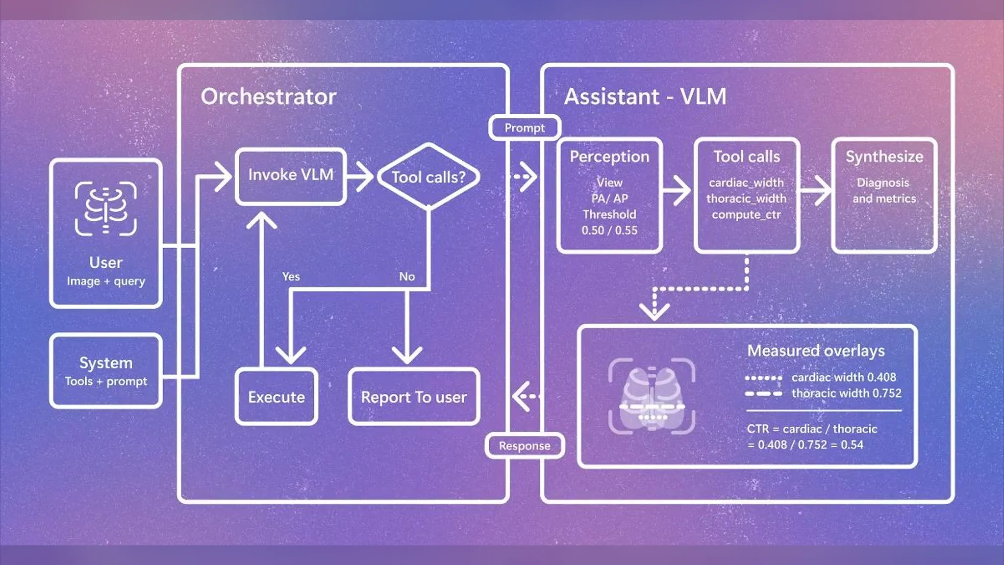
  • The system works by having an orchestrator decide whether tool calls are needed, while a VLM (vision-language model) assistant measures cardiac width and thoracic width to compute the cardiothoracic ratio (CTR)
  • In the example image, a cardiac width of 0.408 and thoracic width of 0.752 were measured, yielding a CTR of 0.54, which falls within the 0.50–0.55 clinical threshold range used to determine cardiomegaly
Video from the source
발표
Microsoft Research, 2026년 8월 11일 X 게시
시스템명
CARE-X
핵심 결합 요소
유연한 추론 + 보정된 예측 + 측정 기반 도구
구조
Orchestrator(도구 호출 판단) + Assistant-VLM(지각·도구 호출·종합)
도구 함수
cardiac_width, thoracic_width, compute_ctr
예시 계산값
심장 폭 0.408 / 흉곽 폭 0.752 → CTR 0.54
적용된 임계값
0.50 / 0.55

Two rulers drawn over an X-ray

In one diagram released by Microsoft Research, two measurement lines are overlaid on a chest X-ray image. One measures the widest point of the heart, and the other measures the full width of the thoracic cavity. Dividing the two figures yields the cardiothoracic ratio (CTR) — a long-standing clinical indicator of how much of the thoracic cavity the heart occupies, used to gauge whether the heart is abnormally enlarged. On August 11, Microsoft Research introduced a system applying this approach, called CARE-X, via its official X account.

In the example released, a cardiac width of 0.408 and a thoracic width of 0.752 were measured, producing a CTR of 0.54. In clinical practice, a ratio exceeding 0.5 is typically treated as a suspected finding of cardiomegaly, and this figure sits right at that boundary. Rather than delivering a diagnosis as a sentence alone, the system displays the underlying numbers on screen as supporting evidence.

이미지: X — 프론티어랩

Two layers of interpretation: orchestrator and VLM

CARE-X's structure is divided into two layers. When a user inputs an image and a question, the orchestrator receives it and passes a prompt to the assistant VLM (vision-language model, which processes images and text together). The assistant first perceives the imaging orientation (PA/AP, posteroanterior or anteroposterior), and if it determines a tool call is needed, it executes functions such as cardiac_width, thoracic_width, and compute_ctr. Once these results return to the orchestrator, if no further tool calls are deemed necessary, it compiles a final report and delivers it to the user.

StageHandled byProcessing
InputUserImage + query
DecisionOrchestratorCalls VLM, branches on whether a tool is needed
PerceptionAssistant-VLMDetermines imaging view, applies 0.50/0.55 thresholds
MeasurementTool callscardiac_width, thoracic_width, compute_ctr
SynthesisAssistant-VLMPresents diagnosis together with figures

Organizing the measured values into a table makes the gap from the thresholds clear as well.

ItemValue
Cardiac width0.408
Thoracic width0.752
Calculated CTR0.54
Applied thresholds0.50 / 0.55

From generating reports to verifiable evidence

Until now, most AI systems for chest X-rays have worked by looking at an image and writing out an entire report as a single piece of text. The problem is that it's difficult to trace back what evidence produced those sentences. What CARE-X emphasizes, as reflected in the phrase "measurement-based tools," is a structure that leaves behind computable intermediate values before a diagnosis is made. This means that instead of a physician receiving only a report and judging whether it's right or wrong, they can also check concrete figures — cardiac width and thoracic width — along with the calculation process. Also notable is the way the orchestrator branches on its own whether a tool call is needed. Rather than forcing the same calculation on every image, this suggests the system has built-in flexible reasoning that calls measurement tools only when necessary.

The trend of medical AI moving beyond text generation to leave behind structured evidence isn't unique to CARE-X. Google Research also announced on August 11 that it had added real-time voice and video consultation capabilities to AMIE, its medical AI research system. This too was an attempt to capture non-verbal cues — such as a patient's facial expressions or breathing — that text-only conversation would have missed. While the approaches differ, both cases share a common focus: rather than AI simply producing an outcome, the emphasis is on how to leave behind and make verifiable the process behind its judgments.

So what actually changes

This post alone doesn't confirm how accurate CARE-X actually is or what dataset it was validated against. Still, the direction this structure points to is clear. If AI can present a calculation process — "cardiac width 0.408, thoracic width 0.752, CTR 0.54" — instead of a sentence like "the heart appears enlarged" when reading a chest X-ray, medical staff gain room to recalculate that judgment or review it against different criteria. For hospitals or research institutions looking to adopt reading-assistance AI, how the "evidentiary structure" is designed — rather than just the "result" — is likely to become the next evaluation criterion.